
 Book an appointment
Retinal detachment
Description:
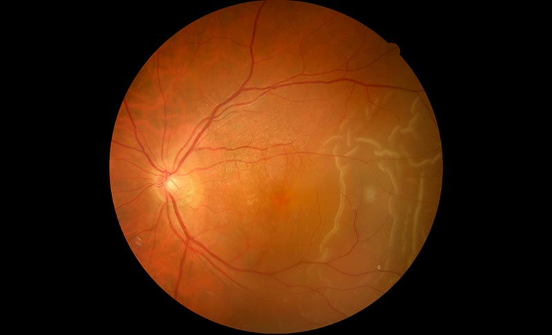
Fundus photography showing a retinal detachment
Symptoms:


- Flashes of light caused by a pulling pressure applied on the retina
- Apparition of floaters in the visual field, indicating intraocular haemorrhage caused by retinal tears.
The area where the retina is detached is not functional. This results in a shortening of the visual field and a partial loss of eyesight, gradually leading to complete blindness when the detachment becomes total and involves the entire retina.
The main risk factors are:
- Myopia
- Eye trauma
- Previous retinal detachment on the other eye
- Cataract surgery
- Some eye inflammations
Treatment:
When the stage of retinal detachment is reached, the only possible treatment is surgery. The surgery is a matter of emergency and is performed most of the time under loco-regional anaesthesia.
The surgery is 20 to 90 minutes long.
Depending on the specificities of each detachment, a variety of surgical techniques may be used: either external surgery, stitching up tears by cryotherapy, or by internal surgery proceeding to a vitrectomy.
Usually, towards the end of the surgery, the surgeon inserts a gas bubble in the eye in order to hold the two retinal membranes together during the time needed for healing.
A few days after the intervention, depending on the localisation of the detachment within the retina, patients must observe a certain positioning of their head.
The gas resorbs gradually within 1 or 2 weeks. Travelling by plane is forbidden before it has totally disappeared.
In some very severe cases of retinal detachment, it might be necessary to inject silicone oil in the eye to ensure the re-attachment of the retina. This silicone oil is usually removed a few months after the intervention.
Post-surgical evolution:
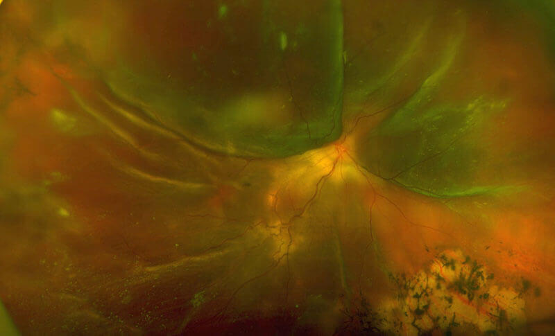
Image of retinal detachment in wide-angle retinography
Post-surgical treatment includes eye-drops and potentially a specific positioning of the head.
Working, sports and driving activities are inadvisable for a duration of 15 to 20 days.
Detachment recurrences are always possible. They require further surgical treatment.
In a very limited number of cases, it is not possible to re-attach the retina because of its rigidity and its tendency to retract.