 Book an appointment
Age-related macular degeneration (AMD)
Description:
AMD is the first cause of legal blindness (visual acuity inferior to 1/10) in France for people above 50 years old.Without treatment, this pathology can lead to the irreversible destruction of the central vision area called macula. The peripheral retina always remains unaffected, which leaves the patient a limited capacity of movement and relative autonomy.
Download the brochure
Symptoms:


Two clinical forms of AMD exist, which might appear separately or jointly:
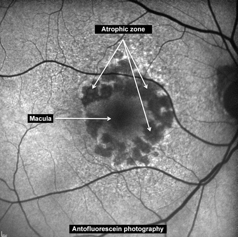
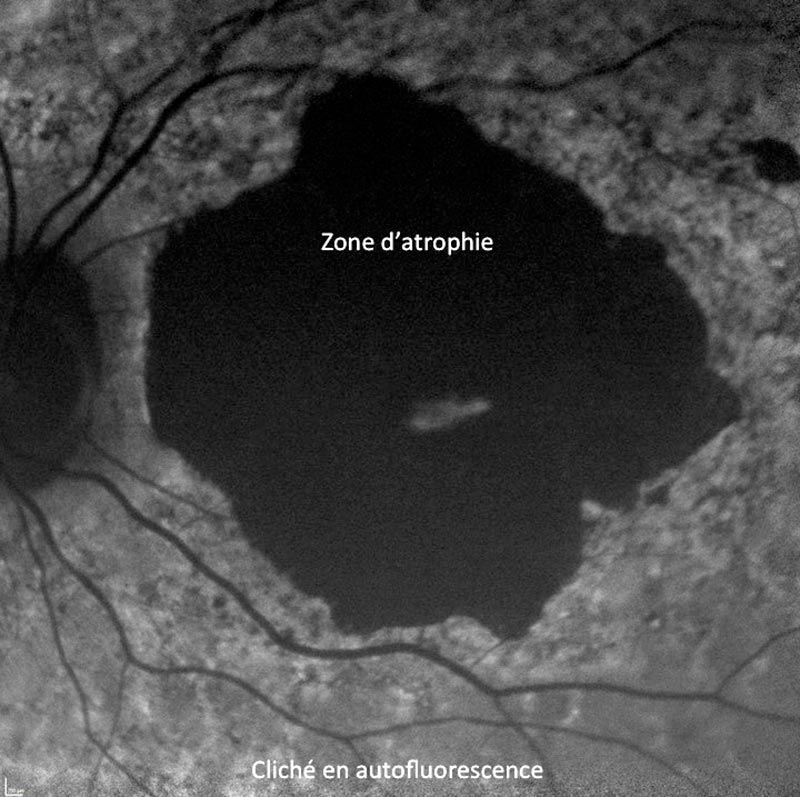
It is characterized by a gradual and irreversible atrophy of the central retina, leaving the patient with only a limited vision for a long time. This form of AMD has a slow evolution but has no efficient treatment outside of lutein and omega-3 dietary supplements which allow to slow down its progression. Quitting smoking and wearing sun-filtrating glasses also allow to diminish its likelihood and to slow down its progression.
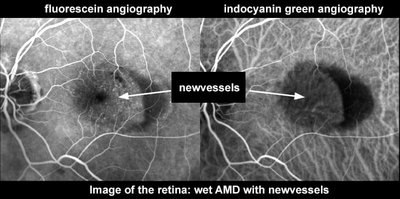
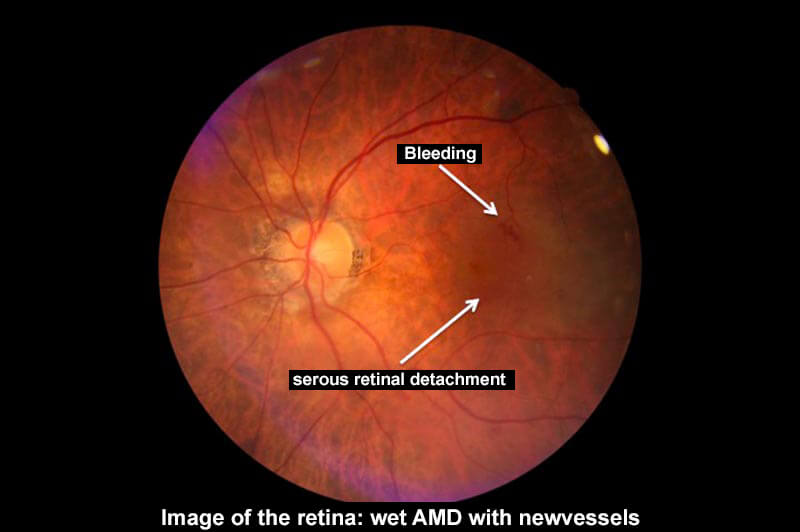
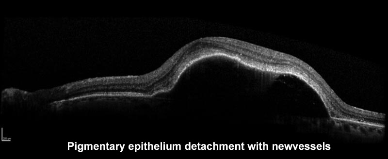
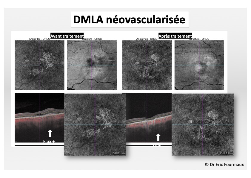
It is caused by the development of abnormal blood vessels under the central retina, themselves caused by the aging of retinal and sub-retinal structures. These new vessels grow fast. Their permeable structure to the liquid components of blood is responsible for the accumulation of these liquid components under or within the retina. The vessels can also break and generate bleeding under the retina.
The diagnosis relies on a fundus examination, OCT examination as well as a fluorescein and ICG angiography. It is important to mention to your doctor any allergy you might have before proceeding to the examination.
Treatment:
The treatment uses a pharmaceutical molecule of the anti-VEGF class. This drug is administered by intravitreal injection. It aims at cauterising the abnormal vessels to stop the liquid flow under and within the retina, in order to increase visual acuity. The cauterization of the vessel itself can sometimes impede on visual acuity.These injections are administered at the Rétine Gallien practice in a room dedicated to this purpose and adapted to its requirements. It is an ambulatory treatment. Prior to the injection, patients will be administered a local anaesthesia and eye-disinfection. The injection is made with a very thin 30 gauge needle, on a very specific spot which poses no risk for the eye. The injection is almost painless and only lasts for a few seconds.
A first series of 3 injections within 1 month is prescribed. In most cases further injections are necessary, at a pace specific to each patient in order to sustain the increase in visual acuity, given the condition has a time-bound evolution.
The most frequent incidents which can arise following this injection include the arising of a subconjunctival haemorrhage (the white of the eye), which is without gravity and naturally resorbs within days; or the perception of small black dots in the visual field in the 24hours following the injection, which are due to the presence of sterile air within the injected product.
Very exceptionally, ocular hypertonia (temporary loss of vision after the injection) can arise and must be referred to your doctor. Intraocular infections, intravitreal haemorrhage or retinal detachment can occasionally arise.
In case you’ve experienced recently (less than 3 months ago) a myocardial infarction or a cerebral infarction, it is important to mention it to your surgeon who will try to find with you the best timing for the injections.
In some particular cases of AMD, other treatments can be suggested such as verteporfin photodynamic therapy (PDT) or vitreo-retinal surgery in case of abundant bleedings under the retina.