
 Book an appointment
Eye tumours
Description:
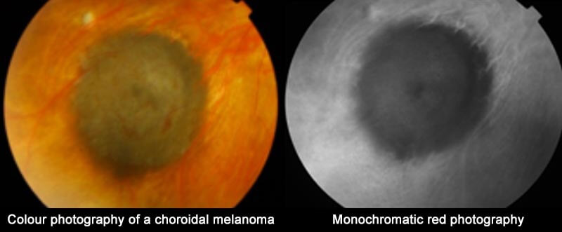
The melanoma is diagnosed with a fundus examination.
Symptoms:
- Flashes
- Floaters
- Reduction of the visual field
- A fall in visual acuity
Uveal melanomas look like a tumour in relief, most often with a pigmented appearance.
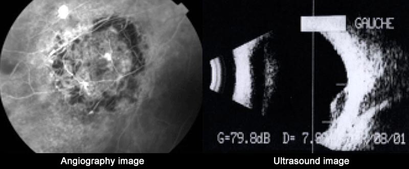
Further examinations of various sorts (mostly angiography and ultrasonography) will confirm the diagnosis and help choosing the best treatment.
Treatment:
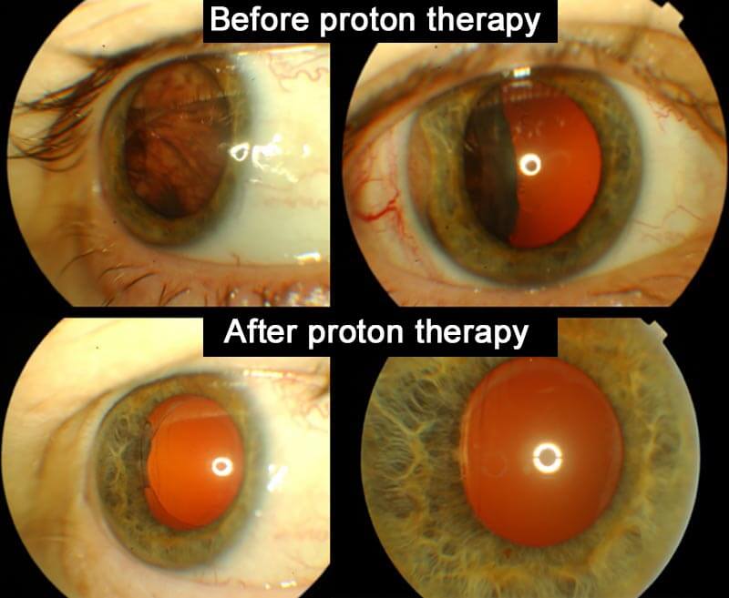
Treatment is variable depending on the melanoma’s localisation and size:For small and medium-sized lesions, we will use conservative therapy (proton therapy, curietherapy and/or exceptionally localised exeresis surgery). However, enucleation remains necessary for major lesions.
Post-surgical evolution:
After exeresis surgery, the eventual complications arise sooner and comprise intravitreous haemorrhage or retinal detachment.
A long and rigorous medical follow-up will be necessary, with general (hepatic ultrasonography) and ophthalmological examinations every 6 months.
Even with a satisfying response to the eye tumour, we observe the development of metastasis in a small number of cases. We can then proceed to chromosomal analysis of the tumour, through a genomic study after transscleral puncture or transvitreal biopsy using 27G TDC cutter.
Most frequently observed anomalies are monosomy3 and multiplication of 8 at the level of tumour cells.
There is a correlation between these anomalies and the patient’s vital prognosis.
New research is being done to identify other molecular markers more specific to a high relapse risk. These molecular signatures could also serve as targets for new therapies.