
 Book an appointment
Retinal vascular occlusions
Description:
Central Retinal Vein Occlusion (CRVO)
ranch Retinal Vein Occlusion (BRVO)
It can be either an occlusion of the central retinal vein (CRVO) or of the branch retinal vein (BRVO), the former often being a more serious condition than the latter which only affects one retinal area among many.
Preventive care rests on identifying risk-factors and dealing with the complications of vein obstructions.
Indeed, there is no known technique to repermeabilise veins. However, retinal hemodynamic can spontaneously improve after an acute episode of developing collateral veins.
Until this technique is set up, treatment of RVO rests on prevention and treatment of potential retinal ischemia complications and of potential macular oedema.
Oedematous complications remain one of the major stakes of RVO.
Treatment:
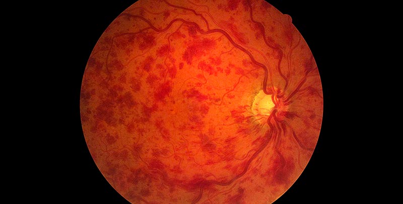
Fundus photography: CRVO with numerous retinal haemorrhages

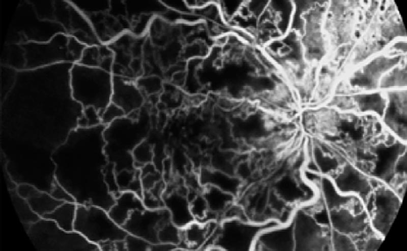
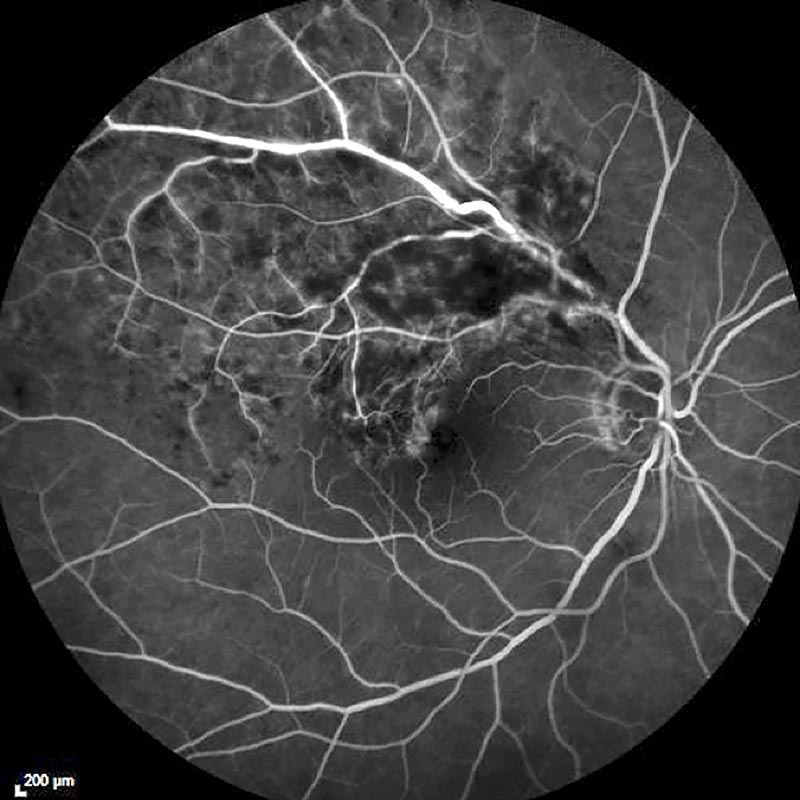
Fluorescein Angiography photography: dilated and tortuous veins
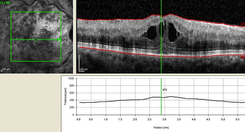
OCT photography: macular oedema
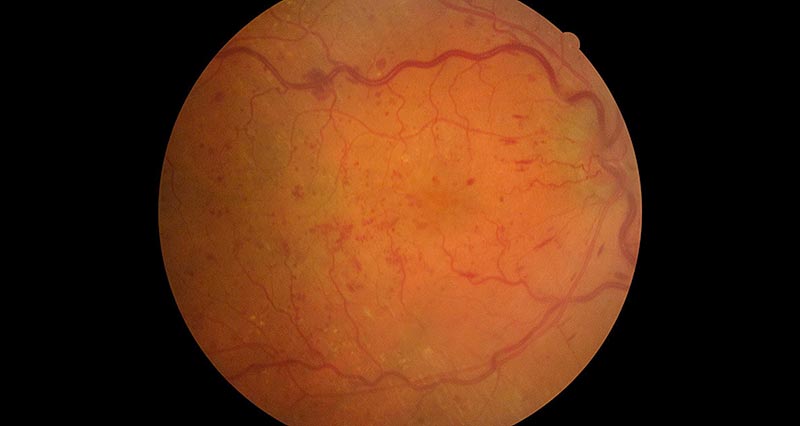
Fundus photography: CRVO with tortuous veins
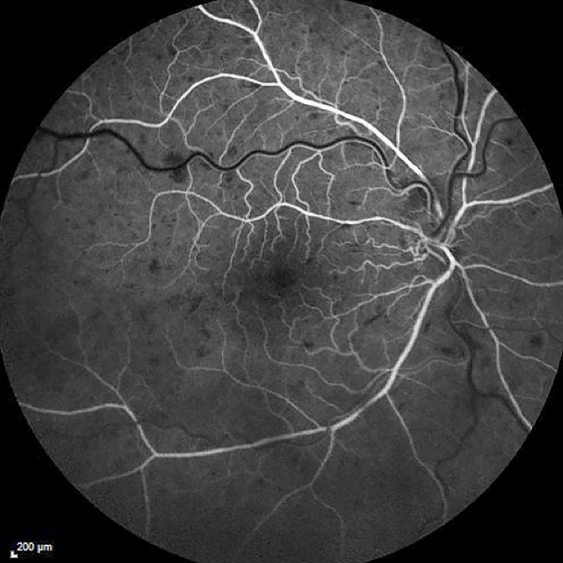
Fluorescein Angiography photography: belated veinous return
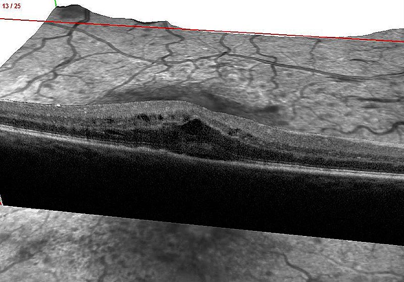
OCT photography: macular oedema
As far as the eye is concerned, a close medical follow-up is key, especially during the first year, while an angiography will look for a potential retinal ischemia and an OCT examination will make it possible to eliminate a potential macular oedema.
Retinal ischemia is treated by laser photocoagulation, which prevents an evolution into a neovascular glaucoma. Multispot lasers make this panretinal photocoagulation possible within 1 to 2 sessions.
This treatment can be paired with intravitreous injections of anti-VEGF.
A macular oedema is treated early with anti-VEGF drugs or corticoids administered in intravitreous injection.
A local photocoagulation might be suggested in a second phase in the case of BRVO.
The prognosis for the patient’s eyesight has considerably improved with corticoids and anti-VEGF injections, and visual acuity is efficiently improved in more than 50% of cases.